|
30 Jun-2 Jul 2021 Montpellier (France)
|
|
|
|
KeynoteUsing morphometrics to understand developmental shifts underlying diversity by Joan T. Richtsmeier
Abstract: Using morphometric approaches to localize differences in form to specific morphological traits or cell populations facilitates the formulation of hypotheses about processes that underlie morphological variation. We are studying mouse models for craniosynostosis syndromes associated with mutations in the Fibroblast Growth Factor Receptor (FGFR) gene family. These mutations can result in constitutive activation of the receptor associated with up-regulation of osteogenic differentiation, and these syndromes are characterized in part by dysmorphology of the bones of the anterior cranial vault and early closure of the coronal suture. Our work with mouse models for FGFR-related craniosynostosis syndromes shows that the closed coronal suture is only one of many complex traits associated with these mutations. Morphometric approaches have helped to identify new targets of molecular, cell, and tissue research in these mouse models for human disease. For example, morphometric analysis of the neonatal mouse skull led us to study cartilages of the embryonic chondrocranium that develops prior to formation of any cranial dermal bone. Our results reveal that FGFR mutations affect the chondrocyte series in addition to the osteoblast lineage, directly affecting embryonic cranial cartilage, a tissue rarely considered in the pathogenesis of craniosynostosis. Understanding the cellular processes that build morphology is key to understanding the disease process and the development of therapies for these syndromes. Our findings are also useful in interpreting processes underlying variation in skull morphology that has emerged over evolutionary time.
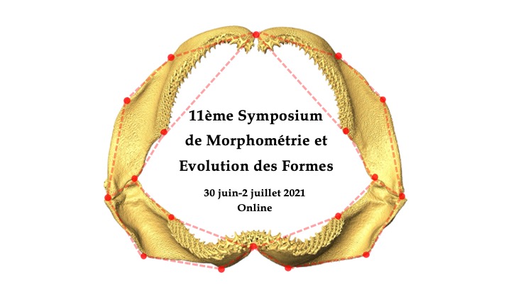
3D reconstruction of micro-computed tomography images of a mouse embryo stained with phosphotungstic acid to reveal the chondrocranium (purple). |


| Online user: 3 | Privacy |


|
